【摘要】 目的:探讨碱性成纤维细胞生长因子(bFGF)与膀胱癌发生发展的关系。方法:应用免疫组化方法检测了48例膀胱癌存档标本和4例正常膀胱组织中bFGF的蛋白表达。结果:48例膀胱癌存档蜡块中有23例阳性,浸润性癌显著高于浅表癌(P<0.01),除G1和G2之间外各分级阳性率之间差异也有显著性。阳性着色定位于肿瘤细胞、血管内皮细胞、正常逼尿肌及肿瘤基质。结论:bFGF可能通过自分泌和旁分泌机制诱导膀胱癌血管生成,或通过胞内分泌作用刺激某些酶的产生,从而促进肿瘤的浸润。bFGF高表达对预测膀胱癌患者的复发和预后有一定的指导意义。拮抗bFGF的作用可能是治疗和预防膀胱癌复发和转移的一条新途径。
中图分类号:R737.14 文献标识码:A 文章编号:1009-4571(2000)06-0604-03
Expression of Basic Fibroblast Growth Factor in Bladder Cancer
BIAN Jia-sheng,FEI Feng-ling, XU zhong-fa,et al.
(Shandong Tumor Hospital, jinan 250117)
【Abstract】 Objective:To study the relationship between basic fibroblast growth factor(bFGF)and development of bladder cancer.Methods:The expression of bFGF protein of 48 samples of bladder cancer kept in archives were detected using immunohistochemical assay with a monoclonal antibody against bFGF.Results:There was no positive signal in 4 normal bladder mucosas. bFGF protein was localized in tumor cells,endothelial cells and normal detrusor muscle. 23 of 48 samples in archive was positive for bFGF protein. There was significant difference between the invasive and the superficial cancers(P<0.01,)the difference also was between the grades except grade Ⅰand Ⅱ.Conclusions:bFGF may induce the angiogenesis of bladder cancer by autocrine and paracrine pathways,or promote tumor invasive by stimulating some enzymes production possibly by an intracrine pathway. Thus antagonizing the action of bFGF may provide an new way in treatment or prevent for the recurrent and matestasis of bladder cancer.
【Key words】 basic fibroblast growth factor (bFGF);bladder neoplasms;immunohistochemical assay;protein
在体外碱性成纤维生长因子(bFGF)是最有效的血管生成因子之一[1],近年来研究表明癌症患者包括膀胱癌患者的尿中bFGF水平是增高的[2,3]。膀胱癌患者的尿具有诱导血管生成的作用,且这种作用已被鉴定部分是由bFGF引起的[4,5]。目前关于膀胱癌组织中bFGF表达的报告很少,bFGF的作用机制也存在争议,bFGF在膀胱癌中表达的临床意义仍不清楚。为此我们应用免疫组化方法对膀胱癌标本中bFGF蛋白的表达进行了检测。
1 材料与方法
1.1 一般资料
选1995年5月~1997年4月同济医院手术切除膀胱癌存档标本48例,其中男36例,女12例;年龄43~81岁,平均64.5岁。按照WHO分级标准,Ⅰ级15例,Ⅱ级20例,Ⅲ级13例,浅表肿瘤33例,浸润肿瘤15例。所选病例均为原发且术前均未接受化疗和放疗。
1.2 免疫组化方法
采用SP法,DAB显色,兔抗人bFGF单克隆抗体滴度1∶50,对每个石蜡标本行4 μm厚连续切片3张,1张重行HE染色观察,1张行免疫组化染色,另1张为PBS替代染色。www.med126.com正常膀胱粘膜为正常对照。抗人bFGF单抗及SABC试剂盒购于武汉博士德公司。
1.3 结果判定
光镜下观察,免疫组化呈黄色或棕黄色者为阳性,每张切片连续观察5个高倍视野,因阳性着色见于肿瘤细胞胞浆、胞核、血管内皮、肿瘤基质、正常逼尿肌,不宜用阳性细胞数作为判断阳性标准,故以平均每个视野中有1处着色即为阳性。

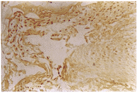
图1 膀胱移行细胞癌I级
(部分癌细胞胞核呈阳性着色,IHC×200)

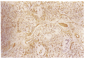
图2 膀胱移行细胞癌Ⅱ级
(部分癌细胞核、间质细胞、血管内皮细胞呈阳性着色,IHC×100)

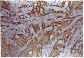
图3 膀胱移行细胞癌Ⅲ级
(癌细胞胞浆阳性、间质细胞阴性,IHC×400)

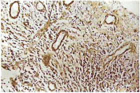
图4 膀胱移行细胞癌Ⅲ级
(血管内皮呈强阳性着色,间质斑状着色,IHC×200)
1.4 统计学方法
统计分析采用SAS软件包,率的比较用卡方检验。
2 结果
免疫组化自身对照片和4例正常膀胱粘膜为阴性,48例存档膀胱癌标本23例阳性,阳性着色见于肿瘤细胞胞浆、胞核、血管内皮、肿瘤基质、正常逼尿肌(图1~4)。免疫组化结果与肿瘤分级和生长方式的关系见表1、2。
表1 48例膀胱癌标本bFGF蛋白表达与肿瘤细胞分级的关系
| 分级 | n | bFGF蛋白(+) | bFGF蛋白(-) |
| G1 | 15 | 3 | 12 |
| G2 | 20 | 9 | 11 |
| G3 | 13 | 11 | 2 |
注:P>0.05(G1 vs G2);P<0.01(G1 vs G3);P<0.05(G2 vs G3);P<0.01(G3vsG1+G2)。
表2 48例膀胱癌标本bFGF蛋白表达与肿瘤生长方式的关系
| 生长方式 | n | 阳性 | 阴性 |
| 浅表 | 33 | 10 | 23 |
| 浸润 | 15 | 13 | 2 |
注:P<0.01。
3 讨论
bFGF不仅是多种细胞(包括上皮细胞、间质细胞和神经元细胞等)的有效有丝分裂原[6~8],还影响许多其他细胞功能,如蛋白酶的产生、细胞潜移和血管生成等[9,10]。近年来的研究表明,bFGF 与膀胱癌的恶性潜能之间有着密切关系。膀胱癌患者尿中可检测到 bFGF 样蛋白,浸润性膀胱癌细胞中也可检测到 bFGF 及其受体 mRNA 和蛋白,而非浸润性细胞株则未能测及[11]。www.med126.comO′Brien等人发现 bFGFmRNA在正常膀胱组织中表达率为87%,而在膀胱癌组织中为12.5%。bFGF蛋白表达在正常膀胱粘膜组织与浸润性膀胱癌之间无差异,但明显高于浅表膀胱癌。bFGF主要定位于正常移行上皮的基底层、正常逼尿肌和肿瘤内血管的部位,而肿瘤细胞少见[12]。本组结果显示 bFGF 蛋白在肿瘤中表达形式多样,肿瘤细胞的胞浆、胞核、血管内皮、平滑肌均有阳性表达,而且不是标本表达形式不一样,有的肿瘤细胞胞核呈强阳性表达,有的仅见于胞浆。高分级浸润性肿瘤 bFGF 蛋白表达明显高于低分级浅表肿瘤。
O′Brien 等依据其实验结果指出,如果 bFGF 不同膀胱癌血管生成的主要因素,那么另外的调节机制参与比肿瘤细胞的直接合成更重要。侵袭性肿瘤侵入基底膜和渗入到膀胱的肌层时, bFGF 便从基底膜细胞和平滑肌细胞等结构中释放出来,刺激肿瘤浸润边缘的血管生成[12]。某些体外实验结果也证实,bFGF 与血管内皮细胞外基质内的氨基葡聚糖形成复合物,此复合物可保护 bFGF 不被水解,从而使 bFGF 以失活形式高量储积。在一定条件下由乙酰肝素酶、血浆酶和组织蛋白酶D作用后释放出来[13]。肌肉内的 bFGF 可能也是以这种形式储存的。浸润性肿瘤细胞可分泌多种酶,包括乙酰肝素酶、尿激酶和组织蛋白酶等降解上述宿主结构而造成浸润[12]。本实验在正常的平滑肌和血管外基质中也检测到 bFGF 蛋白的存在,因此间接地支持了这一理论。
虽然O′Brien等并不否认肿瘤细胞也可合成 bFGF ,但却肯定膀胱癌中 bFGF 的主要来源不是肿瘤细胞[12]。Nguyen指出,免疫组化结果在某种程度上可确定血清、尿中增高的 bFGF 的可能来源[2]。由此,我们认为肿瘤细胞也是 bFGF 的重要来源。这一结论与肾细胞癌中 bFGF 的表达也是一致的[14]。
根据以上结果推测,bFGF 可能通过自分泌和或旁分泌途径促进肿瘤细胞的增殖、血管生成和浸润。这一观点得到了体外实验的支持[15]。事实上,有15%~30%浅表膀胱癌可能进展为浸润性膀胱癌。有研究表明,bFGF 可通过胞内分泌途径调节膀胱癌细胞基质金属蛋白酶的产生,从而增强膀胱癌细胞系的侵袭能力[16]。本组结果发现,低级、浅表型膀胱癌有30.3%(10/33) bFGF 阳性表达,而且肿瘤细胞核阳性主要为低级的浅表型膀胱癌。这是否与 bFGF 通过胞内途径调节膀胱癌细胞基质金属蛋白酶的产生有关,尚需增加观察例数以及进一步的研究。
参考文献:
[1] Rifkin D,Moscatelli D.Recent developments in cell biology of basic fibroblast growth factor[J].J Cell Biol,1989,109:1-6.
[2] Nguyen M,Watanabe H,Budson A,et al.Elevated levels of the angiogenic peptide basic fibroblast growth factor in urine of bladder cancer patients[J].JNCI,1993,85:241-242.
[3] O′Brien TS,Smith K,Cranston D,et al.Urinary basic fibroblast growth factor in patients with bladder cancer and benign prostatic hypertrophy[J].Bri J Urol,1995,76:311-314.
[4] Chodak G , Haudenschild C , Gittes R , et al . Angiogenic activity of
amarker of neoplastic and preneoplastic leisions of the hum an bladder[J].Ann Surg,1980,192:762-771.
[5] Chodak G,Hospelhorn V,Judge S.Increased of fibroblast growth factor-like activity in urine from patients with bladder or kindey cancer[J].Cancer Res,1988,48:2083-2088.
[6] Basilico C and Moscatelli D,The FGF family of growth factors and oncogens[J].Cancer Res,1992,59:155-164.
[7] Tanaka A,Miyamoto K,Minamino N.et al.Cloning and characterization of an androgen-induced growth factor essential of the androgen-dependent growth of mouse mammary carcinoma cell[J].Proc Natl Acad Sci USA,1992,89:8928-8932.
[8] Miyamoto M,Naruo K,Seko C,et al.Molecular cloning of a novel cytokine cDNA encoding the ninth member of the basic fibroblast growth factor family which has a unique secretion property[J].Mol Cell Biol,1993,13:4251-4259.
[9] Miyake H,Hara I,Yoshimura K,et al.Introduction of basic fibroblast growth factor gene into mouse renal cell carcinoma cell line enhances its metastatic potential[J].Cancer Res,1996,56:2440.
[10] Folkman J and Klagsbrun M.angiogenic factors[J].Science,1987,235:442.
[11] Allen LE and Maher PA.Expression of basic fibroblast growth factor and its receptor in an invasive bladder carcinoma cell line[J]. J Cell Physiol,1993,155:368.
[12] O′Brien T,Ganston D,Fuggle S,et al.Two mechanisms of basic fibroblast growth factor induced angiogenesis in bladder cancer[J].Cancer Res,1997,57:136-140.
[13] Saksela O,Moscatelli D,Sommer A,et al.Endothelial cell-drived heparam sulfale binds basic fibroblast growth factor and protects it from proteolytic degradation[J].J Cell Biol,1998,107:743-751.
[14] Eguchi J,Nomata K,Kanda S,et al.Gene expression and immunohistochemical localization of basic fibroblast growth factor in renal cell carcinoma[J].Biochem Biophys Res Comm,1992,183:937-944.
[15] Hori A, Sasada R,Matsutani E,et al.Supression of solid tumor growth by immunoneutralizing monoclonal antibody against human basic fibroblast growth factor[J].Cancer Res,1991,51:6180.
[16] Miyake H, Yoshimura K,Hara I,et al. Basic fibroblast growth factor regulates matrix metalloproteinases production and in vitro invasiveness in human bladder cancer cell lines[J].J Urol,1997,157:2351-2355.

