Anatomical structures involved
The most important structures involved are the lungs and the pleurae and it is essential to understand the normal positions of these. If you cannot remember, go back to the thorax tutorial and check.
Click here to return to the thorax tutorial and revise lung positions.
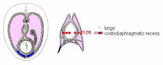
Fluid is likely to accumulate in the costodiaphragmatic and costomediastinal recesses. It is therefore these places which are aspirated during thoracocentesis; this is for several reasons. www.lindalemus.com
1) They present a large area in which there is no viscera, hence fluid can collect here freely.
2) Because the lung or other structures do not extend into these recesses, thoracocentesis performed here is safer since there is a low risk of lung laceration or damage to other structures. 
Figure 44 - Diagrams to show the important recesses in thoracocentesis
Unlike the lungs, the pleural cavities potentially occupy most of the extent of the thoracic cavity. However, as described, this space is reduced by the visceral structures.
DORSALLY
At the boundaries parietal pleura is reflected such that its two faces are directly applied to each other. Caudal to the basal border of the lung this arrangement occurs as costal pleura on the chest wall is reflected off the diaphragm forming the line of COSTOPLEURAL REFLECTION. This is important as it encloses the costodiaphragmatic recess and limits the cranial extent of the pleural cavities. The size of the recess varies with the level of section due to the convex projection of the diaphragm caudally.
CRANIALLY
Cranially the costal and mediastinal pleura dome to form CUPOLAE PLEURAE which extend past the cranial thoracic aperture.
DEVIATION OF PLEURAL CONTOUR
On the right hand side the vena cava is enveloped by a ventro dorsal extension of the pleura - THE PLICA VENAE CAVAE.
Thin pleura may allow communication between left and right sacs.
SUMMARY
Pleura are more extensive than observed from thoracic boundaries due to numerous folds. However, on the dorsal diaphragm there is a small space in which the pleura do not lie.
HORSE
The costodiaphragmatic reflection runs from the eighth rib cartilage to the vertebral end of the seventeenth rib. It slopes, is dorsocranially concave, and is projected cranially at the last rib.
The right cupola pleura extends medially.
RUMINANTS
The costodiaphragmatic reflection runs from the eighth costochondral junction to the twelfth rib, below the lateral margin of the iliocostalis. It has a steep caudal ascent and crosses the middle of the eleventh rib.
The right cupola pleura extends cranially, 2-3cm costally. Medially it regions the mediastinum.
CARNIVORES
The costodiaphragmatic reflection runs from the eighth costal cartilage to the dorsal end of the thirteenth rib. It passes the costochondral junction of the eleventh rib.
Both right and left cupolae pleurae extend, the left further than the right.
PIG
The costodiaphragmatic reflection runs from the seventh costochondral junction to the dorsal half of the last rib. It is a uniform curve.
N.B. in all species the costodiaphragmatic reflection runs from the sternum then rises at the described location e.g. eighth rib.
Clinical note:
In all species the needle must be inserted cranial to the costodiaphragmatic line or else the needle will enter the abdominal cavity.
Clinical note on the ribs:
Although the ribs are merely bypassed by the needle during thoracentesis it is necessary to know their structure for several reasons:
1) The operator should know about the prescence of intercostal spaces as it is through these that the needle is inserted. Since it is usual to insert the needle through a specific intercostal space every time the technique is performed it is useful to know the number of ribs and certain landmarks over ribs. The olecranon is over the 5th rib in large animals and the pig but over the 4th rib in small animals.
2) The presence of the caudomedial groove on the rib containing the neurovascular bundle and the needle inserted to avoid this. Hence, preferably in the middle of the intercostal space or near the cranial edge of the rib.